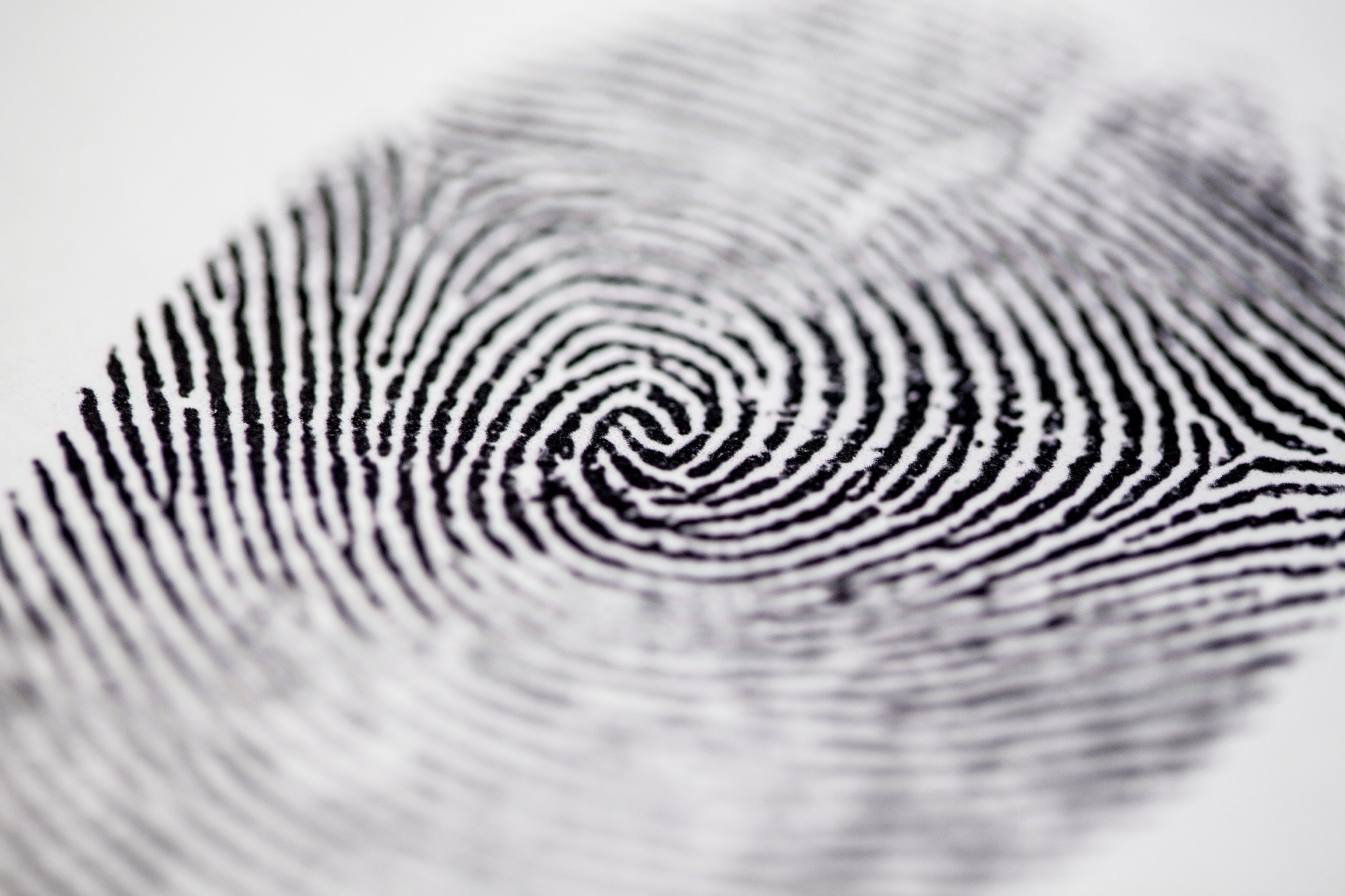
A fingerprint is both complex and unique, and that is why it has fascinated scientists for so long. New research reveals the details relating to their formation and how their characteristic variability is achieved during embryonic development.
Study: The developmental basis of fingerprint pattern formation and variation. Image Credit: Derek Hatfield / Shutterstock
Introduction
Not much is known about how fingerprints form in their intricate patterns. Fingerprints are found on the skin on the palmar side of the fingers and the corresponding side of the toes. They add friction to the skin, aiding in improved grip but also helping to distinguish textures better.
On the fingers, the first two-thirds of the skin is seen to carry transverse ridges. The skin over the terminal phalange, however, displays complex patterns that have been used for over two centuries to establish an individual’s identity. The patterns most frequently seen include arches, loops, and whorls, but others, like tri-radii, are also observed.
However, the factors determining how the primary ridge pattern forms remain unclear. The current study, appearing in the journal Cell, explores the underlying basis of fingerprint development in a unique fashion in every individual.
What did the study show?
Fingerprints are epithelial outgrowths or ridges in which the development of hair follicles is cut short.
This occurs due to the altered development of epithelial placodes because of EDAR and WNT signaling. The placodes do not recruit mesenchymal cells and therefore fail to form hair follicles, unlike the skin in the rest of the body. Fingerprint ridges, therefore, lack markers of late hair follicles.
Thus, epithelial appendages share a common early set of markers, followed by divergence as they move apart.
The researchers found that in the embryo, fingerprint ridges appear early. They begin at the center and apex of the terminal phalanx and move apart as the finger grows. These ridges do not appear to result from overcrowding and mechanical deformation of the epithelium but are produced autonomously by the epithelium itself rather than based on any external driver.
The WNT and EDAR pathways, counterpoised by BMP pathways, develop a Turing reaction-diffusion system via a network of signals. This drives high epithelial growth in localized regions, forming bands of proliferating epithelium in the basal epithelium, overlaid by a thickened suprabasal layer that has undergone unusually rapid differentiation. This forms the fingerprint ridges.
“Meeting this condition of a soft proliferative basal epithelium under a stiffer overlying suprabasal canopy is sufficient to explain ridge downgrowth in the volar epithelium.”
The earliest epithelial ridges to become fingerprints may be seen by about the 13th week of gestation on the fingertip pads. Next, ridge formation progresses over the palmar skin, followed by sweat gland formation from the thickest parts, to be completed by week 17. Smaller ridges then form in between the primary ridges but do not grow downwards, unlike the latter.
The elevated skin above the primary ridges bears an array of sweat gland pores at the very top, as sweat glands begin to grow down once the ridge has reached its maximum depth.
The pattern is selected by week 15, indirectly dependent on the length of the fingers and the shape and size of the fingertip pads, set by around this time. The spaces between consecutive ridges in fingerprints are determined by interactions between WNT and BMP signaling. The latter inhibits WNT activity and ridge formation.
The pattern itself, of swirls, loops, and circles, is due to the initiation of ridge formation from different sites on the palmar aspect of the fingers.
The initiation sites are determined by these signaling pathways as well as the anatomical characteristics of the fingers themselves. From these sites, waves of epithelial proliferation occur that spread outwards until they meet other waves from adjacent points.
“The propagation and meeting of these waves [determine] the type of pattern that forms.”
Thus, fingerprint pattern formation occurs via a dynamic system beginning at multiple sites separated in space. The palm and finger creases do not bear epithelial ridges due to inhibition by changes caused by the markedly thinner suprabasal layer at these sites, coupled with their constant movement.
 Study: The developmental basis of fingerprint pattern formation and variation. Image Credit: Giuseppe Flandoli / Shutterstock
What are the implications?
“Relying on a dynamic patterning system triggered at spatially distinct sites generates the characteristic types and unending variation of human fingerprint patterns.”
The patterning system is responsive to the anatomic features of the fingers, including the boundary separating the volar from the dorsal skin, the volar pad’s size and shape, and the flexion creases. This is added to by random differences and the development of the sweat glands.
Further research may uncover other drivers and inhibitors involved in this system and understand the formation of the fingerprints and related features of the skin, with medical as well as forensic significance.
