Today we are surrounded by artificial chemicals, both those introduced deliberately and those that contaminate the environment inadvertently as a consequence of their use in other applications.
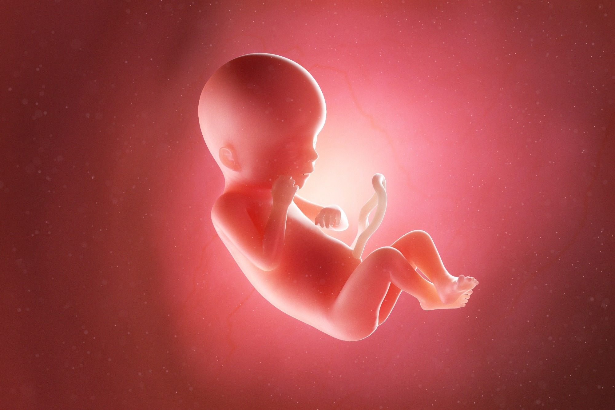
Endocrine-disrupting chemicals (EDC) are of special concern, especially when exposure occurs in fetal life. A new study looks at fetal growth rates related to mercury exposure.
Introduction
Many EDCs are obesogens, including mercury, which has been reported to be correlated with metabolic syndrome. Recently it has come to light that the blood mercury levels in women living in South Korea are about 4.5 g/L, which is quite high relative to the 0.65–1.35 μg/L and ~9 ng/g reported in women in the USA and Japan, respectively.
Mercury exposure is primarily via fish consumption, especially in the form of methylmercury (MeHg). This is an organic mercury compound that builds up in fish flesh. When ingested by pregnant women, it can cross the placenta to accumulate in the fetus.
The Korea Ministry of Food and Drug Safety has set limits on fish consumption in pregnancy because of this, including fish like mackerel and cod, of which 400 g is the upper limit per week, as well as shark and tuna, with a recommended intake of only 100 g per week.
In the current study, published in Environmental Research, the researchers used a physiologically-based pharmacokinetic (PBPK) model to calculate the predicted concentration of MeHg in a given organ with time. It does so using the substance’s pharmacokinetics (the absorption, distribution, metabolism, and elimination [ADME]) and the intensity and route of exposure.
This model can also help estimate the integrated exposure dose. The mathematical prediction of the amount of Hg in the pregnant woman’s body allows for reverse dosimetry, leading to an estimate of the internal dose of the chemical.
The researchers sought, in this study, to find the burden of Hg in the fetus due to transplacental absorption and how this affected fetal growth. The data came from the Children’s Health and Environmental Chemicals in Korea (CHECK) study that began in January 2011 and ended in December 2012.
This included approximately 330 pregnant women attending several university hospitals in South Korea with their newborn infants at the time of birth. Blood and urine samples from pregnant women and the umbilical cord and placental samples were tested for MeHg, along with the first urine and meconium. In addition, expressed breast milk and the infant’s hair were collected on day 30.
The measured Hg level was applied to the model, and the exposure amount was calculated after adjusting for fetal growth and an increase in maternal blood, richly-supplied tissues, and fat compartments during gestation. The amount of Hg passing through the placenta to collect in the fetal plasma was calculated to arrive at the fetal body burden of Hg.
What did the study show?
The geometric mean (GM) birth weight was 3.3 kg and 3.2 kg for boys and girls, respectively. The GM Hg concentration in maternal and cord blood Hg was ~4.5 and ~7.4 μg/L, respectively, measured in just over a hundred paired samples. The former is like the human biomonitoring-1 (HBM-1) value and indicates that Hg exposure needs to be moderated during pregnancy in the individual concerned.
In contrast, placental and meconium concentrations went up to 9.0 and 36.9 ng/g, respectively. Infant hair samples (n=25) showed a GM of ~440 ng/g. Therefore, Hg levels were higher in meconium and cord blood compared to maternal blood, corroborating the findings of earlier studies.
Fetal tissues, including placenta, cord blood, meconium, and infant hair, are all enriched for Hg compared to maternal blood, with hair samples showing concentrations 20-174 times that of maternal blood.
While 95% of the mothers had blood Hg concentrations below 8.7 μg/L, the corresponding level in 95% of neonate cord blood samples was 17.2 μg/L. In proportion, the MeHg level in cord blood was estimated at 13.4 or below for 95% of samples. In contrast, only 5% of cord blood samples had MeHg levels below 4.
Overall, Hg levels were lower in this study than Japanese or Singapore studies suggest but higher than those reported in the USA or Canada.
Therefore, the calculated fetal body burden of MeHg ranged between 26.3 to 86.9 mg in this cohort. Five rounds of follow-up assessed postnatal Hg concentrations in cord blood, with 75% of values being below 9.6 μg/L.
The exposure during fetal life affected newborn length at birth, which showed a positive correlation with cord blood Hg. This holds good even after adjusting for maternal characteristics, including the body mass index (BMI). However, head circumference and birth showed no such correlation.
Postnatal growth was not statistically associated with cord blood Hg levels. Still, trends for rapidly increasing weight gain were observed in the high-exposure group after six months of life for both sexes. This indicates that apart from the individual constitution, age, weaning practices, and child behavior, Hg levels affect weight gain.
The presence of lead could also influence these findings, with increased length and weight being reported to be linearly related to lead concentrations in cord blood. When lead and Hg exposure were implemented in the mixed model, no relationship was observed with length or weight.
What are the implications?
Earlier studies have been unable to agree on the association of Hg exposure and growth rates, with some reporting an increase and others a decrease. With this study, too, the significance of the presence of Hg in the diet is established, but data as to the nature of its significance remains to be collected.
However, the fact that Hg exposure has an adverse impact on the fetus makes it needful to set upper limits on such exposure during pregnancy.
The Environmental Protection Agency (EPA) has already set a reference dose of 0.1 μg/kg/day for MeHg, sufficient to avoid adverse effects over a lifetime of such exposure.
An earlier study by the same authors showed Hg exposure to be linked to hyperlipidemia and raised liver enzymes, probably due to its ability to inhibit the breakdown of oxidized lipids, that are toxic to the host. This is accompanied, however, by induced oxidative stress and systemic inflammation, influencing the build-up of abnormal fat cells.
Further research is required with specific information, such as fish diet after birth and co-exposure to other environmental contaminants to clarify and generalize the relationship between Hg and growth.”
