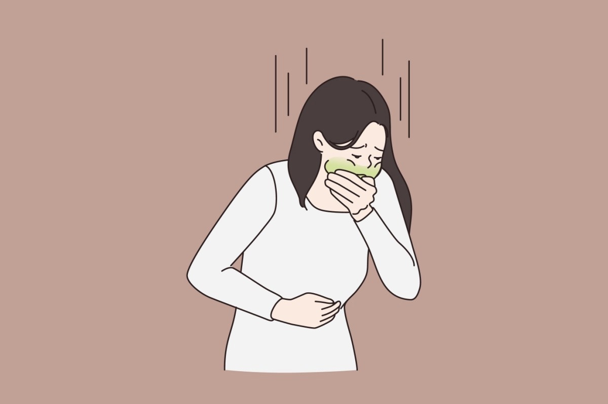
In most cases, the presence of toxins in food can cause nausea and vomiting. These are bodily defenses aimed at minimizing the duration of exposure to the toxin. The pathways by which the brain detects the presence of such toxins and synchronizes various defenses remain poorly understood.
Study: The gut-to-brain axis for toxin-induced defensive responses. Image Credit: Drawlab19 / Shutterstuck.com
A new Cell journal paper describes a system by which gut-brain pathways coordinate with brain circuits to initiate these defensive reactions. This involves a set of nerve cells called Htr3a+, which act on the dorsal vagal complex (DVC) to cause retching and a reflex avoidance of certain flavors.
The study findings indicate that these responses are triggered by both chemotherapy and food poisoning, with these toxins acting through a common set of circuits.
Introduction
Retching and vomiting involve motor responses that are reflexively triggered, though initiated by the brain. These are accompanied by the sensation of nausea, thus helping the individual to identify the toxic substance so that they can avoid it in the future. This phenomenon is known as conditioned flavor avoidance (CFA).
Nausea and vomiting are the most common adverse effects associated with chemotherapy. This has stimulated intense research into the mechanism by which these responses arise. Some studies have suggested a gut-brain axis as the underlying cause of both responses when the body is exposed to an enterotoxin or chemotherapeutic drug.
Vagotomy, as well as using 5-hydroxytryptamine 3 receptor (5-HT3R) and neurokinin 1 receptor (NK1R) blockers, successfully prevent both vomiting and nausea. However, this leaves several questions unanswered, including the specific cells involved, their projections, and molecular signals that mediate this response.
About the study
The current study uses laboratory mice to address such questions. Although mice lack a vomiting response to emetics, they can demonstrate conditioned flavor avoidance and appear to retch, making them a suitable animal model.
Mice were exposed to staphylococcal enterotoxin A (SEA), which causes food poisoning and vomiting. This was found to induce a peculiar mouth-opening response that lasted approximately five times longer, as well as a wider stretch of the jaw, than spontaneous responses. This resembled retching-like behavior and was accompanied by synchronous electromyogram findings of the diaphragm and abdominal muscles.
Even though these are inspiratory and expiratory responses, respectively, they showed simultaneous bursts of activity, unlike the alternating activity typical of normal breathing in mice. Moreover, the diaphragm exhibited more robust and more rapid activity during the phase of opening relative to the phase of closing the mouth during this muscular action, thus supporting the hypothesis that this is a type of retching-like behavior.
SEA also induced CFA in mice, with both CFA and retching reduced by granisetron, a 5HT3R antagonist, and CP-99994, an NK1R blocker. This indicates that SEA acts by circuits involving these receptors.
Study findings
As suggested by earlier research, the scientists found that the vagus nerve mediates vomiting in response to toxins. In addition, cutting the diaphragmatic branches of the vagus nerve on both sides significantly reduced both retching and CFA in mice.
Using genetic labeling methods, a population of Hrt3a+ neurons was identified. These sensory vagal neurons carry signals triggered by toxins when they encounter enterochromaffin cells. These signals eventually reach Tac1+ neurons in the DVC.
Chemogenetic inactivation of DVC neurons caused reduced retching in response to SEA. These findings indicate the presence of a gut-brain axis mediating the SEA-induced retching and CFA.
Over one-third of DVC Tac1+ neurons were activated by SEA. These neurons are known to produce neurotransmitters like glutamate and specific Tac1+-encoded neuropeptides.
Specific Tac1+-encoded neuropeptides bind to NK1R, which is a key emetic signal, thus supporting the theory that these proteins, as well as glutamate, are key to nausea and retching when the animal is exposed to SEA. This was not found with other DVC neurons or other emetics like lithium chloride.
A single-synapse long pathway was found to be directly linked with Tac1+ neurons to specific Hrt3a+ vagal sensory neurons on the same side and several brain areas. These vagal neurons appear to respond to 5-HT from the enterochromaffin cells, the nerve endings of 5-HT being in close proximity to enterochromaffin cells. Moreover, the enterochromaffin cells likely mediate a selective response to SEA.
This Tac1+-Hrt3a+-enterochromaffin circuit forms the gut-brain pathway that mediates defensive nausea, vomiting, and retching in response to SEA. Tac1+ neurons determine how long and intense each retching movement is in response to signals carried by Hrt3a+ vagal sensory neurons in the gut.
Stimulation of these neurons through optogenetic signals led to retching-like behavior in a dose-dependent manner. This was confirmed by chemogenetic activation which led to CFA.
These data suggest that activation of Tac1+ DVC neurons is sufficient to induce defensive responses in mice.”
DVC neurons project to different areas of the brain, depending on their location in the DVC. As a result, different subsets caused selective retching or CFA in response to SEA.
In fact, chemogenetic activation confirmed that each of these responses was selective to a particular subset. These are represented by the Tac1+ DVC-rVRG and DVC-LPB pathways, respectively.
The first of these is possiblly due to the recruitment of respiratory neurons that subsequently leads to retching-like responses. The second may involve CGRP+ neurons that mediate conditioned taste aversion (CTA) learning, thereby causing CFA.
Tac1+ neurons also appear to contribute to chemotherapy-induced retching-like and CFA responses, with the same selectivity of response observed for different neuron subsets following intraperitoneal injection of the chemotherapeutic drug, doxorubicin.
Interestingly, in vitro experiments suggested an indirect activation of the gut-brain circuit by SEA and doxorubicin, as direct contact with these toxins failed to activate nasogastric (NG) cells or enterochromaffin cells. The toxins appeared to act through inflammation induced by them, which causes the release of interleukin 33 (IL-33). This alarmin molecule binds to its receptor on the enterochromaffin cells, thereby causing 5HT release that stimulates vagal sensory cells.
What are the implications?
The current study reports the presence of a gut-brain pathway mediating toxin-induced vomiting and nausea through two different brain circuit systems in mice. By expelling food from the stomach, these responses defend the host from toxins in food.
The existence of Tac1+ cells, which are a subset of DVC cells that are key to these toxin-induced defenses, was revealed. Another subset of cells known as AP neurons may also participate in these responses.
Further studies should examine the reason for residual retching-like behavior after ablation of the diaphragmatic vagal innervation, which could be due to the role of spinal efferent nerves. The effects of ablating multiple genes in the Tac1+ neuron population on toxin-induced defenses also remains to be studied.
